KPV Peptide Research: α-MSH, Inflammatory Signaling & Intestinal Models
What published experimental research has investigated about KPV: its relationship to α-MSH, PepT1 transport, NF-κB and MAPK signaling, and chemically induced intestinal inflammation models.
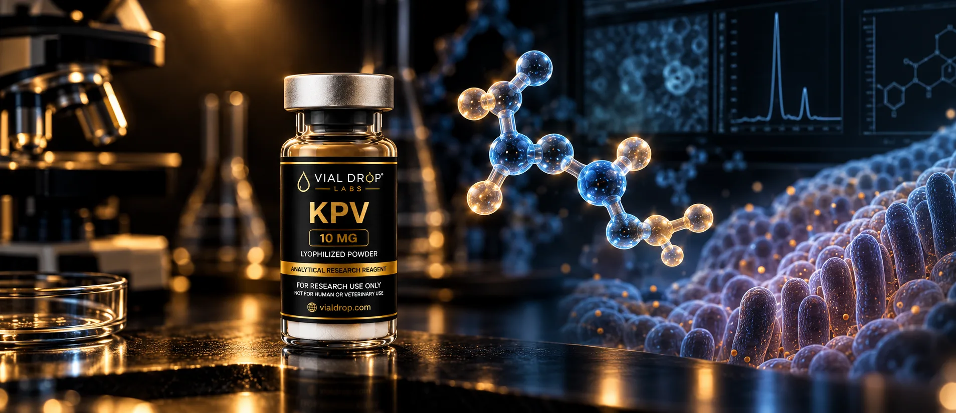
KPV is a remarkably small peptide. Just three amino acids—lysine, proline and valine—make up a molecule that has been used in experimental research spanning inflammatory signaling, peptide transport and intestinal models.
Its size is only part of what makes KPV interesting.
The sequence KPV corresponds to the final three amino acids of alpha-melanocyte-stimulating hormone, better known as α-MSH. That relationship led researchers to ask whether some biological activity associated with the larger melanocortin peptide could be retained within this much smaller fragment.
The answer has turned out to be more complicated than a simple fragment-versus-parent relationship. Studies have examined KPV in cell systems and animal models, investigated how it enters certain cells, and explored whether its observed activity depends on the same melanocortin receptors associated with α-MSH.
For the molecular identity, analytical characteristics and documentation associated with the compound itself, see the VDL KPV Research Reference. This article focuses on a different question: What has published experimental research actually investigated about KPV?
KPV and α-MSH: related by sequence, not identical
α-MSH is a 13-amino-acid peptide derived from proopiomelanocortin. KPV—Lys-Pro-Val—corresponds to residues 11 through 13 at its C-terminal end.
That makes KPV an α-MSH-derived sequence, but it does not make the two molecules interchangeable.
Full-length α-MSH contains structural information that KPV does not. Conversely, isolating the three-residue sequence gives researchers a way to investigate whether certain experimental observations can persist without the rest of the α-MSH molecule.
That distinction became particularly interesting when researchers began examining melanocortin receptor signaling.
A 2003 study by Getting and colleagues compared different α-MSH-derived sequences in a mouse model of crystal-induced peritonitis. Their results suggested that the C-terminal KPV sequence did not behave identically to other portions of α-MSH. The investigators specifically explored whether the observed effects depended on known melanocortin receptors and reported evidence consistent with a different mechanism for KPV.
That does not establish a single, definitive mechanism for KPV. It does, however, help explain why the tripeptide became an independent subject of research rather than simply being treated as abbreviated α-MSH.
Other melanocortin-related compounds illustrate the same need for molecular precision. A synthetic α-MSH analog such as Melanotan II, for example, is a separate chemical entity with its own structure and research profile. A relationship to the melanocortin system does not make these compounds equivalent.
Following the inflammatory signaling pathways
Some of the most detailed KPV experiments have looked downstream at intracellular signaling.
One important target has been nuclear factor kappa B (NF-κB). NF-κB is not a KPV-specific pathway; it is a broad transcriptional regulatory system involved in cellular responses to inflammatory and other signals.
Researchers have also examined mitogen-activated protein kinase (MAPK) pathways, another family of signaling systems involved in how cells respond to their environment.
In a 2008 Gastroenterology study, Guillaume Dalmasso and colleagues exposed intestinal epithelial cell lines and Jurkat T cells to inflammatory stimuli and studied what happened in the presence of KPV. They measured NF-κB activity, MAP kinase signaling and inflammatory cytokine expression using several laboratory methods.
The investigators reported reduced activation of NF-κB and MAP kinase pathways under the experimental conditions they studied, along with changes in pro-inflammatory cytokine secretion.
Those findings are useful because they move the discussion beyond a vague description of KPV as “anti-inflammatory.” They identify specific signaling systems that researchers measured under controlled experimental conditions.
They also need to be read within those conditions. Cell signaling experiments can reveal mechanisms worth investigating, but they do not by themselves establish what a compound will do in a person.
PepT1 adds another piece to the KPV story
The same Dalmasso study addressed another question: How does such a small peptide get into certain cells?
Researchers focused on peptide transporter 1, or PepT1.
PepT1 transports small peptides across cell membranes. The researchers investigated whether KPV could interact with this transport system and whether PepT1 participated in its cellular uptake.
They used human intestinal epithelial cell lines and a human T-cell line, along with uptake experiments designed to characterize the transport process. Their results supported PepT1-mediated uptake of KPV in the experimental systems they studied.
That observation connected two parts of the research: transport and signaling.
Rather than simply exposing cells to KPV and measuring downstream changes, the investigators had evidence for a pathway by which the tripeptide could enter cells in their models and then examined changes in NF-κB, MAP kinase signaling and cytokine expression.
The PepT1 finding is sometimes presented far more broadly than the experiment warrants. It does not establish targeted delivery in people, nor does it demonstrate clinical effectiveness. It describes a transport mechanism observed under defined experimental conditions.
The distinction becomes even more important when moving from cultured cells to animal models.
From cultured cells to intestinal models
Dalmasso and colleagues extended their work into two established mouse models of intestinal inflammation.
One used dextran sulfate sodium (DSS). The other used 2,4,6-trinitrobenzene sulfonic acid (TNBS).
These models create intestinal injury and inflammatory responses experimentally. Researchers can then examine tissue changes, inflammatory markers and other biological responses under controlled conditions.
In the Dalmasso study, KPV was evaluated in both DSS- and TNBS-induced mouse colitis. The investigators assessed outcomes that included histological inflammation and expression of inflammatory cytokines. They reported differences in inflammatory measures in the KPV experimental groups.
A separate study by Kannengiesser and colleagues also examined KPV in murine intestinal-inflammation models. That work evaluated KPV in DSS- and TNBS-based experiments and further investigated the role of melanocortin signaling in the observed responses.
Together, the studies provide a useful progression of experimental evidence: molecular signaling, cellular transport and animal models.
They do not, however, turn a chemically induced mouse model into evidence that KPV treats inflammatory bowel disease in people.
That distinction matters.
DSS- and TNBS-induced colitis are research tools. They allow investigators to reproduce selected features of intestinal inflammation in animals and test biological hypotheses under controlled conditions. Crohn's disease and ulcerative colitis in humans are far more complex.
Results in these models can justify additional research. They cannot establish clinical efficacy.
Does KPV work through melanocortin receptors?
This may be one of the more interesting unanswered questions in the KPV literature.
Because KPV comes from the C-terminal sequence of α-MSH, an obvious hypothesis is that its observed biological activity should depend on melanocortin receptors.
Experimental results have made the picture less straightforward.
The Getting study compared the core and C-terminal regions of α-MSH and investigated receptor involvement in their observed effects. The authors reported evidence suggesting that KPV's activity in their experimental model did not depend on the same melanocortin receptor pathway examined for other α-MSH-derived sequences.
The later intestinal work also investigated melanocortin receptor involvement rather than simply assuming that KPV reproduced α-MSH signaling.
That leaves an important distinction:
Sequence origin is not the same thing as mechanism.
KPV can be accurately described as the Lys-Pro-Val sequence corresponding to the C-terminal end of α-MSH. That chemical relationship is clear.
It does not follow that every experimental observation involving KPV must result from conventional α-MSH receptor pharmacology.
The literature instead points toward a more nuanced research question involving transport, intracellular signaling and potentially receptor-independent or partially receptor-independent mechanisms.
That's a much more useful scientific question than forcing KPV into a single pathway before the evidence supports doing so.
KPV research isn't limited to intestinal models
The intestinal literature receives considerable attention, but KPV has also been investigated in other cellular systems.
In 2012, Stephen Land examined KPV and other melanocortin-related peptides using immortalized human bronchial epithelial cells.
The study looked at inflammatory signaling triggered by TNF-α and respiratory syncytial virus-related stimuli, with particular attention to NF-κB signaling and chemokine secretion. The work also investigated IκBα stabilization and nuclear movement of the NF-κB component p65RelA.
This provides another experimental setting in which researchers explored KPV's relationship with inflammatory signaling.
There is an important wording distinction here as well: human-derived cells are not a human clinical study.
Experiments performed in human cell lines can provide information about cellular mechanisms in a human biological context. They cannot establish clinical efficacy, safety or therapeutic outcomes in living people.
What does the evidence actually tell us?
Looking across these studies, a coherent research picture begins to emerge.
KPV has been investigated as a discrete tripeptide rather than merely as shorthand for α-MSH. Researchers have examined its uptake through PepT1, measured its relationship with NF-κB and MAP kinase signaling, evaluated cytokine responses, tested it in chemically induced mouse models of intestinal inflammation, and investigated whether its experimental activity requires conventional melanocortin receptor signaling.
That's a meaningful body of mechanistic and preclinical work.
But the type of evidence matters just as much as the findings.
The studies discussed here consist primarily of:
- controlled cell-culture experiments;
- experiments involving human-derived cell lines;
- mechanistic signaling studies; and
- murine models of experimentally induced inflammation.
Each can answer particular research questions. None substitutes for controlled human clinical evidence.
These experimental findings do not establish clinical efficacy or therapeutic benefit in humans. They also do not establish human dosing, clinical safety or effectiveness for inflammatory bowel disease, IBS or other digestive or inflammatory conditions.
The value of the literature is more specific: it identifies biological mechanisms and experimental observations that can be tested, challenged and developed through additional research.
Published biology and analytical identity answer different questions
There's another distinction that's easy to miss when reading peptide research.
A published paper can tell us what researchers observed when they studied a compound they identified as KPV.
It cannot tell us what's inside a completely different vial.
Those are separate scientific questions.
Published biological research deals with experimental models, mechanisms and observations. Analytical testing deals with the identity and characteristics of a particular research material.
For an individual lot, questions such as identity, purity and measured content require analytical evidence tied to that material. A citation to a KPV study cannot substitute for that documentation.
Vial Drop Labs maintains its available analytical reports in the Research Documentation Library. Researchers evaluating those reports may also find our guides to reading a certificate of analysis, HPLC versus mass spectrometry, and purity versus content per vial useful for understanding what different analytical measurements actually establish.
Keeping those evidence streams separate makes both easier to interpret.
The published literature tells us why scientists have studied KPV.
Analytical documentation tells us what was measured in a particular research material.
Neither should be used to claim more than it demonstrates.
Sources
- Getting SJ, Schiöth HB, Perretti M. Dissection of the Anti-Inflammatory Effect of the Core and C-Terminal (KPV) α-Melanocyte-Stimulating Hormone Peptides. Journal of Pharmacology and Experimental Therapeutics. 2003;306(2):631–637. PMID: 12750433. DOI: 10.1124/jpet.103.051623.
- Dalmasso G, Charrier-Hisamuddin L, Nguyen HTT, Yan Y, Sitaraman S, Merlin D. PepT1-mediated tripeptide KPV uptake reduces intestinal inflammation. Gastroenterology. 2008;134(1):166–178. PMID: 18061177. PMCID: PMC2431115. DOI: 10.1053/j.gastro.2007.10.026.
- Kannengiesser K, Maaser C, Heidemann J, et al. Melanocortin-derived tripeptide KPV has anti-inflammatory potential in murine models of inflammatory bowel disease. Inflammatory Bowel Diseases. 2008;14(3):324–331. PMID: 18092346. DOI: 10.1002/ibd.20334.
- Land SC. Inhibition of cellular and systemic inflammation cues in human bronchial epithelial cells by melanocortin-related peptides: mechanism of KPV action and a role for MC3R agonists. International Journal of Physiology, Pathophysiology and Pharmacology. 2012;4(2):59–73. PMID: 22837805. PMCID: PMC3403564.
Stay ahead of the research
Get new research articles, product updates, and subscriber-only offers delivered to your inbox.
See our Privacy Policy.
Educational content is provided for laboratory research understanding only. It is not medical advice and does not describe or endorse any use outside legitimate research settings.